EBUS-TBNA: a practical referral guide for GPs
CLINICAL REFERRAL GUIDE · 13 SEPTEMBER 2026
EBUS-TBNA: a practical referral guide for GPs
When mediastinal or hilar lymphadenopathy warrants specialist assessment, how EBUS-TBNA can support diagnosis and staging, and what to include in a referral.
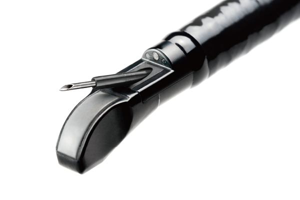
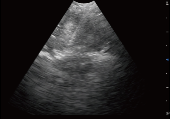
The role of EBUS-TBNA
Endobronchial ultrasound-guided transbronchial needle aspiration combines bronchoscopy with real-time ultrasound to sample accessible lymph nodes and lesions adjacent to the central airways. In suspected lung cancer, selecting a target that provides both diagnosis and nodal staging can reduce separate procedures. Conventional bronchoscopy alone is not an equivalent test for mediastinal nodal staging.
Which patients may benefit?
- Patients with suspected lung cancer and enlarged or PET-avid mediastinal or hilar nodes, when the result would alter treatment.
- Patients requiring tissue confirmation of otherwise unexplained intrathoracic lymphadenopathy.
- Selected patients with suspected sarcoidosis when tissue sampling is needed; a safer accessible peripheral biopsy site may be preferable.
For potentially curative lung cancer pathways, CT, PET-CT and tissue sampling should be coordinated. A negative EBUS result does not exclude malignancy when suspicion remains high; additional sampling or surgical staging may be required. NICE lung cancer guidance.
Granulomas do not establish sarcoidosis on their own. Alternative diagnoses, including tuberculosis, must be considered; microbiology requirements should be discussed before sampling. Suspected lymphoma may need additional tissue for classification. ATS sarcoidosis diagnostic guideline.
When to consider referral
Refer promptly for suspicious nodes or a lung lesion on imaging, especially with haemoptysis, weight loss or progressive symptoms. Referral can be for diagnostic planning: the GP does not need to select the final biopsy method or arrange PET-CT before contacting the specialist.
Helpful referral information
Include the clinical question, symptom timeline, smoking and TB history, CT report and access to images, previous tissue or microbiology results, relevant comorbidities, oxygen needs and medication list. Highlight anticoagulants and antiplatelets; changes require an individual procedural plan.
Discuss a referral
Referrals are welcome by phone on 044 803 2497 or by email to reception@dredgar.co.za. All referrals are screened to triage urgency, and an appointment will be promptly arranged.
George consultations begin in November 2026. Milnerton consultations continue until the end of October 2026.
Educational information for healthcare professionals. Referral decisions and investigations should be individualised to the patient and local resources. Phone and email referrals do not replace emergency assessment.